

We treat a wide range of conditions affecting the chest, including:
-
Barrett’s esophagus
-
Chest wall diseases
-
Esophageal cancer
-
Esophageal motility disorders, such as achalasia
-
Esophageal stricture
-
Gastroesophageal reflux disease (GERD)
-
Hiatal hernia
-
Lung cancer
-
Lung cancer staging
-
Malignant pleural effusion
-
Mediastinal tumors
-
Mesothelioma
-
Metastasis of other cancers to the lungs
-
Myasthenia gravis
-
Paralysis of the diaphragm
-
Pneumothorax (collapsed lung)
-
Solitary pulmonary nodule
-
Thymic cancer
-
Thymoma
-
Tracheal diseases
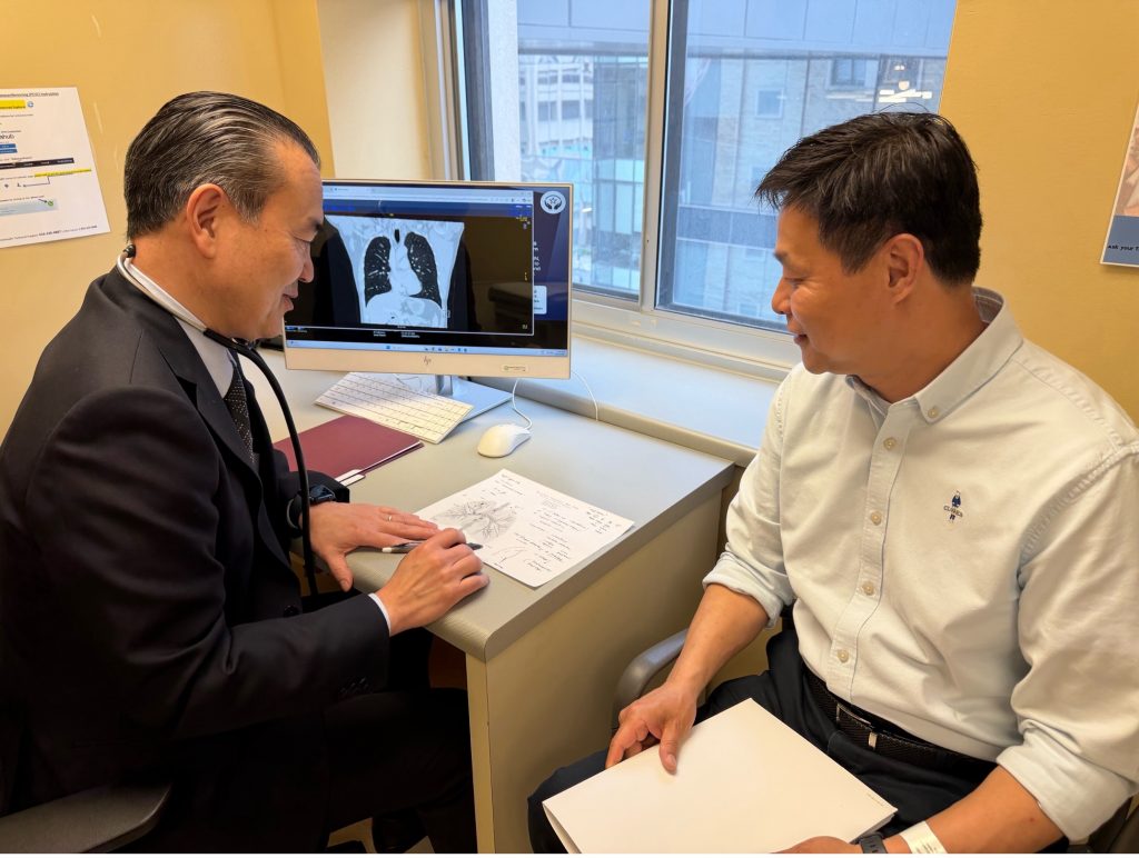
Lung Cancer Rapid Assessment and Management Program (LungRAMP) allows patients who are suspected of having lung cancer to obtain consultation and state-of-the-art diagnostic investigations through a streamlined process. Specialist consultations, tests and, often, a biopsy are typically required to establish a diagnosis. The care we provide you will include some or all of:
-
Pre-consultation
-
Consultation
-
Diagnosis and decision-making
-
Smoking Cessation Intervention, for active smokers
We treat patients with esophageal disorders such as reflux and difficulty swallowing. We investigate for esophageal cancer by conducting a variety of tests to diagnose and stage the disease. We also work closely with Medical and Radiation Oncologists – both locally at Princess Margaret Cancer Centre as well as with your referring oncologists, to provide timely and personalized treatment.
Thoracic surgery is the surgical subspecialty that deals with diseases of the chest (thorax), as well as the trachea, esophagus and stomach. Some of the more common conditions we treat include lung cancer, esophageal cancer, metastatic disease to the lungs and end-stage lung disease from any cause. Many of these conditions are treated using minimally invasive surgery.
Many patients with cancer develop secondary tumours in their lungs or other organs. The spread of cancer from one organ or place in the body to another, non-adjacent organ or body part is called metastasis. When these secondary tumours occur in the lungs only, there may be a possibility to remove or destroy them.
The Division of Thoracic Surgery and the Radiation Medicine Program have developed the Lung Metastasis Program. This highly specialized diagnostic and management service assesses patients with cancer of any primary site other than the lungs where cancer has spread to the lungs (lung metastasis) to assess surgery or radiotherapy as treatments for lung metastasis. Our goal is to prolong life and improve quality of life by offering a multidisciplinary assessment and cutting-edge surgical or radiation therapy treatment in the most efficient and supportive way.
The Rapid Assessment of Complex Pleural Effusion Program (RACE) is a part of the Division of Thoracic Surgery, managing and addressing the needs of patients with Malignant Pleural Effusions (MPE). MPE is a type of advanced malignancy that can cause an accumulation of fluid in the chest. MPE affects nearly 50% of patients with advanced cancer, most commonly patients with lung, breast, gynecological cancers and lymphoma. The goal of the program is to shift the inpatient management of MPE to an ambulatory setting and, by doing so, increase our patients’ quality of life and reduce healthcare costs. The program aims to offer prompt access to clinicians with expertise in MPE assessment and management and long term follow up of patients with MPE in a multidisciplinary team setting.
Tests/Services
Thoracentesis
-
A thoracentesis is a procedure done to remove the extra fluid in the space between your lungs and chest wall. Some conditions cause more fluid to build up in your pleural space and too much fluid can make it more difficult to breathe. A thoracentesis is done to ease the symptoms related to pleural effusions, and the doctor may send a small sample off to the laboratory for more testing.
Tenckhoff Catheter Insertion
-
A Tenckhoff Catheter is a soft, see-through rubber tube that remains within the pleural space and can be permanent if necessary. A doctor puts it into your pleural space to drain fluid from around your lung that allows you to drain the re-accumulating fluid as needed from your home environment to ease symptoms caused by the fluid buildup.
Tenckhoff Catheter Removal
-
The fluid amount in the lung space may reduce significantly and you may no longer require the catheter for drainage. If your catheter is draining minimal amounts (less than 150mL a week) for a period of 3 weeks, chest x-ray done that looks stable, and your symptoms are minimal and/or manageable- then you can come to the clinic for an assessment to see if the catheter can be removed.
Chronic thromboembolic pulmonary hypertension (CTEPH), similar to other types of pulmonary hypertension (PH), reduces blood flow and increases pressure in the blood vessels of the lung. Breaking its name down helps us to understand what CTEPH is.
-
‘Chronic’ means a condition that lasts a long time (months-years)
-
‘Thromboembolic’ refers to the complete or partial blockage of a blood vessel by a blood clot that has broken away from where it was formed.
-
‘Pulmonary’ relates to the lungs.
-
‘Hypertension’ is the medical term for high blood pressure.
CTEPH is a type of pulmonary hypertension that is caused by unresolved or recurrent pulmonary emboli leading to chronic pulmonary vascular obstruction by an organized clot. The disease progresses despite adequate anticoagulation as a result of secondary arteriopathy eventually causing right heart failure and death.
Recognition of CTEPH in patients with a history of pulmonary emboli is important to achieve timely diagnosis and appropriate referral and follow-up.
Patients with CTEPH can present with acute pulmonary emboli. CTEPH should be suspected in the presence of:
-
Idiopathic and/or recurrent pulmonary emboli
-
Longer time between symptom onset and diagnosis
-
Right ventricular systolic pressure > 50 mmHg on echocardiogram
-
Large perfusion defects, mosaic parenchymal changes, and/or arterial webs or bands on CT Pulmonary Angiogram